

To me, to you, the picture might look like nothing special. But Lee McIlwain was struck by it.
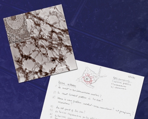
On May 15, 1999, McIlwain sat at his desk flipping through a textbook. Today, he doesn’t remember exactly why he checked out that book from the library. But in it he came across a review of some 20-year-old research by Sheldon Penman, a respected cell biologist. Penman’s work had little to do with McIlwain’s. But McIlwain’s eye was drawn to a picture — an electron-microscope image that showed a human epithelial cell with hundreds of tiny structures crisscrossing inside.
McIlwain realized that if what Penman had seen in these epithelial cells was true, it might explain some things in McIlwain’s work that had seemed a mystery.
McIlwain, professor of cell and molecular physiology, had been studying large nerve cells known as spinal motor neurons for 25 years — about the same amount of time that his lab technician, Victoria Hoke, had been working with him. The two were trying to understand the size and shape of these neurons, hoping that they could provide some insight into Amyotrophic Lateral Sclerosis (ALS) — commonly called Lou Gehrig’s disease.
When McIlwain looked at this image from Penman’s work, he began to see his motor neurons in a whole new way. He read Penman’s papers. By Monday, May 17, McIlwain was handwriting notes — questions, instructions — on the back of used office paper. He also sketched how the insides of spinal motor neurons might look if they contained what he called the “intracellular matrix.” The next day, he was at the whiteboard, hashing out his ideas with Hoke, sketching the matrix again with a red marker.
That one image from Penman’s work made so much sense to McIlwain that he switched gears and began a series of experiments that would consume his and Hoke’s work life for almost two years. If Penman had been right, and epithelial cells contained a framework that held the cells together, then maybe motor neurons did too.
The only trouble was, scientists had never agreed that Penman was right. Scientists had accepted the idea of a cytoskeleton in the nucleus and cytoplasm of cells. But they had reservations about the framework that Penman had isolated. Penman and other scientists debated the idea. But not even Penman, a member of the National Academy of Sciences, nor Keith Porter, another prominent cell biologist and National Academy member, could get scientists to agree.
But McIlwain couldn’t resist. The idea had such “explanatory power” that he had to pursue it.
Before he found that picture, McIlwain had been experimenting with spinal motor neurons’ size and shape. When he would injure a motor neuron by cutting its axon, the cell would swell. But it didn’t just swell. The parts of the cell — the nucleus, the nucleolus, and the cell body — changed size in proportion to one another, and the nucleus moved away from the center of the cell. The cell seemed to have something keeping all its parts in line.
McIlwain and Hoke tried to determine what proteins were increasing during the swelling. They ran gel after gel — gel electrophoresis, a procedure used to separate and identify proteins. “We used two-dimensional gels, thinking that any protein that was really going to be of interest to us was going to be on those gels,” McIlwain says. But they couldn’t find what they were looking for.
Besides running the gels, they would measure the amount of protein in the cell body after cutting the cell’s axon. The protein increased by 50 percent within three weeks after the cell was injured. But the gels didn’t show the increase. “Where is that fifty percent protein increase that should be on these gels?” McIlwain thought. One possibility was that the cells contained a lot of protein that couldn’t be identified using gels. Insoluble protein — protein that can’t be dissolved by the usual detergents.
While McIlwain was stewing over the missing protein, he came upon Penman’s electron microscope image. The picture got him thinking — maybe there were insoluble proteins in motor neurons that formed a matrix similar to the one that Penman had seen.
On his handwritten notes, McIlwain listed experiments he should try. One instruction reads simply, “image it.” McIlwain didn’t know what he was getting himself into.
To view cells with electron microscopy, scientists cut thin-sections — they slice lengthwise through the cell, using diamond knives or plate glass. To get the super-thin slices, scientists use an embedment material, a hard plastic.
But Penman had used a different way of thin-sectioning. Instead of plastic as the embedment material, he used a flexible wax. And he removed the wax before viewing, which allowed him to see an entirely new structure in the cell.
McIlwain needed to do the same thing with his motor neurons. He and Hoke soon realized one reason why this technique is unusual — it’s hard to get it right. The wax can become brittle and hard to work with. Mess up once, and you must start over with a new slice.
Hoke had never done electron microscopy before. She worked at it. The wax kept falling apart. She and McIlwain modified the wax so that it would hold up. Then the slices of cell kept coming out very thick. The thick slices made the pictures too dark.
“At first we got really ugly images,” McIlwain says. “But then, peeping through were some things that looked like we were on the right track,” he says. “So we kept going back.”
Hoke worked some more. “Vickie hardly ever drops an experiment,” McIlwain says. “She is careful and deliberate, and at the end of the experiment, you know that it’s reliable.”
After six months of learning the new technique, and almost two years after McIlwain had discovered that picture, McIlwain and Hoke, together at their department’s electron microscope, finally got their first good images of the matrix structure.
“When those pictures came out, it was just so obvious. We’re talking about looking at the same thing that Penman had been looking at in nonneuronal cells,” he says.
McIlwain was sure that the structure they were seeing was holding the cell together. Before examining the cell, they extracted its soluble protein using 10-normal sodium hydroxide, which “dissolves protein like crazy,” as McIlwain says. The substance had extracted about 60 percent of the total protein from the cell. “You remove half the protein, but you still get something that looks like a cell,” Hoke says. This convinced them that the cell contained insoluble proteins that were controlling its size and shape.
The two also found success with experiments that followed. “For a while, every experiment we did was suggested by Penman’s work, and we could predict how it was going to come out,” McIlwain says. The experiments showed that the size and shape of the isolated framework changed after nerve injury, that the framework contained all the missing protein, and that the framework was very insoluble.
McIlwain has no doubt about the matrix. He’s one of the few. One criticism: when Penman was promoting this idea, some scientists argued that one part of the thin-sectioning method — removing the wax and drying the cells with a technique called critical point drying — could be altering how the fine details of the framework appeared.
McIlwain concedes that could be true. But whatever the fine details of the framework, something is maintaining the cells’ size and shape, even after McIlwain removes the soluble proteins. “I think these are the true bones of these cells,” he says.
The explanation satisfies him enough that he’s willing to spend the next few years — the end of his career — refining this work. “I’m happy to just stand right in line behind Porter and Penman and say, well, I took your ideas and applied them to neurons, and by golly, they seem to check out,” he says.
To agree with McIlwain, scientists will have to change they way they think about cells. Bringing about such a “paradigm shift” is not easy. “It’s hard for scientists to believe that something this fundamental has been overlooked,” McIlwain says. “The whole idea of the cytoskeleton is so familiar that it’s hard for scientists to accept new ideas about it.”
If the proteins in the matrix are indeed insoluble, they’re going to be hard to identify. McIlwain is betting that mass spectrometry — a tool of the new field of proteomics — will help. He has begun sending human cell samples to Carolina’s Proteomics Core Facility. There researchers break the proteins down into their small chains, known as peptides, and run them through a mass spectrometer to determine their mass. Then McIlwain can use the human genome to identify the peptides, giving him a clue as to which proteins they come from.
McIlwain believes that the framework is not made of the usual cytoskeleton proteins. These proteins are soluble, and the way he sees it, the matrix is not. “I may be able to show that there’s some interesting new protein in motor neurons,” he says.
McIlwain hopes that proteomics will help scientists see the “insoluble world” in cells. “This world exists in nonneuronal cells as well as in neurons,” McIlwain says. “We’ve got to come to grips with it.”
“It doesn’t happen very often that you just feel like something comes along and synthesizes what you’re doing,” he continues. “For me the real reward has been in getting an answer that really made good sense.”
This work was funded by two small grants from UNC-Chapel Hill — a University Research Council grant and a Medical Alumni Foundation Endowment grant.