


Jack Griffith picks up a three-inch-high stack of journal articles, the result of a recent literature search. He flips through a few pages. “Gels and plots and tables,” he says. Another paper. “More gels, more gels. And aahk, more gels. It’s pretty dense.”
Griffith, professor in the Lineberger Comprehensive Cancer Center and the Department of Microbiology and Immunology, knows these studies are important, some of them even vital, to his work. He just wishes that they looked as exciting as they are. “If you can see what the molecules are doing and you have a picture of it, it’s easier to understand, and papers tend to make more of an impression,” he says.
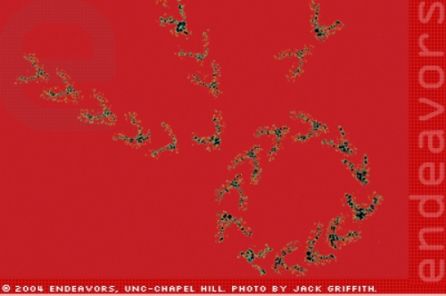
Take for example a journal article that Griffith and Tom Cech, a Nobel Prize-winning biochemist from the University of Colorado, were about to submit in 1995. Cech and Griffith had developed a new method for determining the three-dimensional structure of RNA molecules using Griffith’s favored tool: the electron microscope. They used the method to show the structure of a particular ribozyme (a cell particle that produces proteins). But at first glance, the microscope images looked pretty plain. Black and white shots of the ribozyme — a y-shape. Griffith wanted something better.
“This technique had not been done before,” Griffith says. “Both Tom and I were excited about the work, and of course, we wanted to get it seen by as many people as possible.”
So Griffith spent days putting together an image. “This was before the days of Photoshop,” he says. “I actually cut out each of these ribozyme images by hand and arranged them into what looked to me like a fun pattern.” He taped them to a poster board and photographed the whole thing with black and white film. Then he took it into his darkroom and used color filters and cibachrome paper to make a print showing ribozymes in formation on a red background. The paper made the cover of the October 2, 1995 Embo Journal.
Griffith isn’t afraid to admit that he likes pretty pictures. He takes photos of everything — his greyhounds and horses and Maine Coon cats, a flower covered in snow outside his office. He appreciates anything visual. As he talks, he jots down key words and draws sketches. He wears nice shoes.
Griffith also gets a charge out of all the tinkering that goes along with electron microscopy. “I like the garage, I like big machines,” he says. In his spare time, he restores vintage Jaguars.
But the electron microscope is not a toy. It has become Griffith’s own brand of science, perfected since his graduate student days when he first began building microscopy equipment. His work has led to clues about DNA replication that might have stayed hidden if the microscope hadn’t enabled him to see what was going on, then analyze it. And over the years, Griffith and his lab have used their knowledge of electron microscopy to help other scientists see, too.
“I would describe him as one of the best high-resolution electron microscopists in the world,” says Paul Modrich, professor of biochemistry at Duke University. “His work has addressed problems in DNA replication, repair, and recombination.” Modrich has known Griffith since the 1970s, when Modrich was a graduate student at Stanford University and Griffith was a postdoctoral researcher in the lab next door. Modrich has since collaborated with Griffith on studies of DNA mismatch repair.
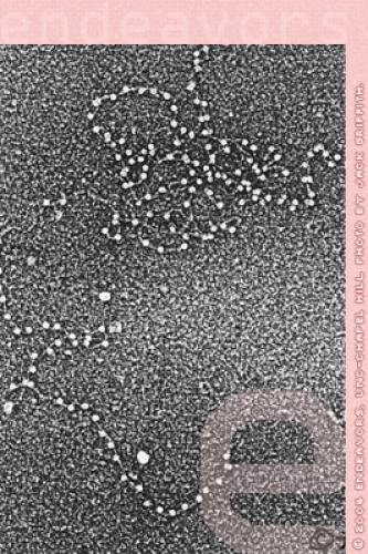
Griffith started working with electron microscopy in the late 1960s, when he was a Ph.D. student at California Institute of Technology. In the mid-1950s, scientists had begun using a technique called metal shadow-casting. Inside high-vacuum machines, metal with a low melting point was heated until it evaporated, then it was sprayed over the raised sample (a virus or whatever the scientist was trying to view). Because it’s slightly elevated, the sample gets coated with more of the metal than the supporting film, and the outline of the metal coating is what the microscope will “see.”
But this technique doesn’t work for viewing something as small as DNA. Scientists had looked at DNA with electron microscopy before, in the early 1960s, by coating it with a thick layer of protein. “So now this twenty-angstrom thick DNA had been made two hundred angstroms thick, which you could see with the electron microscope easily,” Griffith says.
But because the DNA was covered in the thick sheath of protein, scientists couldn’t see DNA’s finer details. At Caltech, Griffith worked in a lab studying chromosome structure. They needed to see not only DNA, but the proteins that are bound to it.
So Griffith started building microscopy equipment and experimenting with shadow-casting, but using a different metal — tungsten. Before, scientists had used metals such as gold and platinum that have a low melting point and are readily evaporated. “But it turns out that the lower the melting temperature, the larger the globs of metal that coat the sample,” Griffith says. “And the larger the globs of metal, the worse the resolution is.” Tungsten, with its higher melting point, produced a finer spray and more detailed image. In his 1969 dissertation, Griffith describes this shadow-casting method and another technique he developed — a way of mounting DNA on very thin carbon films.
Using these methods, Griffith created some of the first electron microscope images showing the basic subunit structure of chromosomes. In 1971, he published a paper with Arthur Kornberg, a Stanford scientist who won the Nobel Prize in 1954 for identifying DNA polymerase, an enzyme responsible for DNA replication. That paper includes the first electron microscope image of DNA bound to a known protein. The work showed that electron microscopy had the potential to do more than take pictures; it could be used to quantitatively analyze DNA.
Today, Griffith’s lab is continually refining its brand of electron microscopy and using it to study the swirling activity of DNA in complex with proteins. He describes his quantitative approach to electron microscopy as “doing biochemistry with a microscope.” He says, “Biochemistry is extremely important. It defines everything that’s going on.” But there are limitations. You can’t look at large pieces of DNA. And it’s hard to find out exactly what is binding where at a particular moment. “When you run these reactions in the test tube, there are so many things going on,” Griffith says. “In general, maybe twenty-five percent of the molecules are engaged in doing what they’re supposed to be doing. So a biochemical study that looks at just the whole average is really unable to go in and say, ‘okay, when the molecule’s replicating perfectly, are there two molecules of polymerase or not?’ Because you can’t see.”
So Griffith’s way is to first do single-molecule electron microscopy — taking high-resolution pictures of a particular DNA structure or an instance of protein binding. “And then you make it biochemically relevant by counting,” he says. “You sit at the electron microscope and scan around fields of these molecules.” A researcher would typically scan one thousand molecules and click a counter each time they find what they’re looking for. Then they can do the math to figure out how common that shape or binding pattern is.
In 2002, Griffith and colleagues used similar quantitative techniques to map the DNA involved in Fragile X syndrome, a developmental disorder. It has been known that in people with Fragile X, a particular DNA sequence is repeated too often — as many as two thousand times, compared to only seventeen to thirty times in normal DNA. But it wasn’t known how that repetition, called expansion, contributed to Fragile X syndrome.
“We showed that in Fragile X, that expansion creates a segment of the chromosome that is very unorganized and unprotected relative to the rest of the chromosome,” Griffith says. The work provides a clue to the molecular causes of the disorder.
Griffith’s lab also uses electron microscopy as a guide in building models of large DNAs in complex with binding proteins. He calls this technique “starting with this pile of parts and pieces and seeing what you can put back together.” Graduate student Nicole Fouche, for example, is building a model of the end of a human chromosome, called a telomere.
“This technique allows you to manipulate things,” Griffith says. “You can add in histones and see what effect they have.” Then add in another protein, and so on. “So you understand, stepwise, the role of each of these proteins by kind of slowly building the house back up,” he says.
“And of course,” Griffith says, surrounded by dozens of framed journal covers, “if you have some clues from even a fuzzy picture, that helps a lot.”