


You drive to your x-ray appointment in a hybrid car equipped with a global positioning system (GPS). As you walk in the door of the hospital, you check your email using your camera phone. But when it’s time for your x-ray, you step up to a mammoth, doughnut-shaped machine that uses technology that hasn’t changed much in a century.
Physicist Otto Zhou plans to bring x-rays into the age of cell phones and GPS by using something called carbon nanotubes. These tiny tubes, made of a single layer of carbon atoms, can help make x-ray machines work faster, come in smaller packages, and produce clearer images.
How can a little tube do all that? Mostly because of its super-small size. “A nanotube is basically as small as you can get in nature,” Zhou says. They can be as small as one nanometer — one billionth of a meter — in diameter. To compare, one human hair is as much as a hundred thousand times thicker. Materials this small have different physical and electrical properties than regular-sized stuff.
Zhou first studied carbon nanotubes just after he finished his doctoral degree in 1992, when he began working as a researcher at Bell Labs, experimenting with nanotubes as a possible substitute for the carbon electrode in rechargeable lithium batteries. Then he moved for one year to the materials research lab at NEC, the Japanese company where nanotubes had been discovered in 1991. “That’s really where I learned all about nanotubes,” he says.
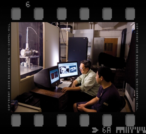
Then Zhou came to Carolina, with nanotubes still on his mind and x-ray machines becoming a part of his daily life. Zhou is a materials scientist — he makes all sorts of new materials — and he would often use x-rays to determine the exact properties of his creations. “X-ray testing is actually one of the most useful techniques to identify or characterize materials,” he says. Zhou compares making new materials to cooking; when you start out, you know basically the ingredients you want to use, but you don’t know the exact proportions. So, you make a test batch, taste it, then revise the recipe. In Zhou’s work, an x-ray machine was a routine part of the taste test.
But with the traditional x-ray machines Zhou was using, the metal filaments that produce the x-rays burned out often because of the high temperatures required to operate them. The lab replaced a tube about once every year, and it wasn’t cheap — six thousand dollars or more each time.
“That’s really where the original motivation was,” Zhou says. “Can I use a nanotube as part of that x-ray source? Hoping it would have a longer lifetime.”
In a conventional x-ray machine, the metal filament emits electrons when exposed to very high heat (a thousand degrees Celsius). Those electrons bounce off a separate piece of metal, generating x-rays. If Zhou could replace the filament with a nanotube, he wouldn’t need to use the scorching heat.
Because nanotubes have such a small diameter, Zhou says, they emit electrons easily, when exposed to just a tiny amount of voltage. Low-voltage would equal low-temperature.
It took some trial and error for Zhou and collaborators to figure out the precise design. Other researchers have worked on producing an x-ray source using “cold” electron sources before. But not carbon nanotubes. “Because of the inferior properties of these early emitters, they were not able to achieve the x-ray intensity that is required for practical use,” Zhou says. Zhou and his collaborator Jianping Lu, also a professor of physics at Carolina, make a breakthrough by demonstrating that a nanotube-based field emission x-ray source can generate enough x-ray intensity for practical applications.
In 2000, Zhou and Lu filed the first patent application on their technology using nanotubes as an x-ray source, and that same year Zhou cofounded a company, Xintek, to develop and commercialize the technology, after licensing it from the Office of Technology Development (OTD) at UNC-Chapel Hill.
“We’re proud of our relationship with Xintek,” says Jim Deane, a project manager at OTD who has worked with Zhou. “As a licensee, they are successfully filling our number-one mission, which is to create new products and make their benefits available to the public.”

In 2002, Zhou, Lu, and Sha Chang of the Department of Radiation Oncology published a report about their first prototype machine, which can operate at room temperature. The lower temperature allows the x-ray source to last longer than the old metal filaments. And because there’s no heat, the nanotube machine is free of bulky insulation or ventilation components, so it’s much smaller than conventional machines. The smaller size would be especially useful for ambulance drivers or industrial inspectors who need a portable x-ray machine, Zhou says.
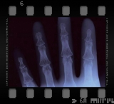
Other advantages of the nanotube-based machine: it needs no warm up or cool down time between images, so it can take them in fast succession. The old metal filaments need time to warm up, like a space heater you might use at home. But the nanotube x-ray source is like an overhead light; you flip the switch, and it immediately comes on. Because of this fast switching time, “You can use the nanotube x-ray almost like a high-speed camera. In fact, it is a high-speed x-ray camera,” Zhou says. So the machine can capture clear pictures of fast-moving objects such as a beating heart. “Because we can take an image very fast, you basically freeze the motion,” Zhou says. The result is like any movie; a succession of still images that, when shown one right after another, create a motion picture.
Etta Pisano, director of the Biomedical Research Imaging Center at Carolina, hopes to work with Zhou to take advantage of the stop-motion ability of the nanotube x-ray machine for use in breast-cancer screening. Any woman who’s had her chest squished in a mammography machine knows that with current x-ray machines the breast must be compressed. The compression makes the image sharper by limiting motion. But Pisano hopes that Zhou’s technology can make compression unnecessary. “Once the x-rays are made faster, the exposure time can be much shorter and we may not have to compress the breast at all, or as much, to get a sharp picture,” she says.


Many researchers have been interested in using nanotubes to improve medical computer tomography (CT), which is used to provide more detailed information on head injuries and brain diseases, for example, than simple x-rays can. In traditional CT scanning, the x-ray source mechanically rotates around objects or patients to collect as many as a thousand two-dimensional images. A scanner uses those to construct a three-dimensional picture.
Zhou has collaborated with Lu, Lin, and Lalush to develop a micro-CT scanner using the carbon nanotube x-ray tube. The scanner is now being used by medical researchers at UNC-Chapel Hill for in vivo cancer imaging in small animal models. Because of the fast switching time of the carbon nanotube x-ray source, the device can provide real-time image of an object in motion.
Earlier this year, Zhou and colleagues reported that they had created a prototype nanotube x-ray machine that can take multiple images from different angles without having to move the x-ray source. Zhou’s experimental machine uses many tiny nanotube pixels that are programmed to fire in fast succession, taking images from different angles while staying put. Zhou’s machine makes the whole process faster and the x-ray machine smaller. A similar machine could also be used in airport and other security scanning.
“If it works as well as we think it will, other advantages will be that scanners will be cheaper, use less electricity, and produce higher-resolution images,” he says. Zhou’s report on this prototype appeared in the May 2005 issue of Applied Physics Letters. Earlier this year, Zhou, Lu, and a former graduate student received the first U.S. patent on the concept of a nanotube-based stationary CT scanner.

Zhou believes he’s found a successful recipe for using nanotubes to make your future x-ray appointments faster and more accurate — soon. “We are working with manufacturers to put the first generation of carbon nanotube x-ray machines on the market for industrial inspection within a year and for use in medical imaging in three to five years,” he says. With support from the U.S. Department of Homeland Security, UNC-Chapel Hill and Xintek are now developing the next generation airport baggage scanners using the carbon nanotube x-ray technology.
Zhou’s startup company isn’t alone in exploring applications for nanotubes. Industry giant Samsung, for example, is pursuing the use of carbon-nanotube emitters in flat-panel displays for computers and TVs. Xintek has teamed up with some companies in Asia to develop this product as well. And to develop the x-ray applications for nanotubes, Xintek is working with several large companies in the security, medical, and industrial inspection businesses.
Though it’s a young company, Xintek is holding its own. In 2004, Xintek won a Technology Innovation Award from consulting company Frost and Sullivan, which cited Xintek’s model of creating products tailored to specific customer needs. A recent market report by NanoMarkets listed Xintek as one of the world’s top twenty firms in the nanotube electronics business.
Back in the lab, Zhou and his team keep working. “I do the research, I cook this material, I bring it out and look at it, test it,” Zhou says. “And then we go back and cook again.”
Randy Mayes was formerly a staff contributor for Endeavors.
Zhou is the Lyles Jones Professor of Physics and Materials Sciences and was the founding director of the N.C. Center for Nanoscale Materials, one of the largest federally funded research centers on carbon-nanotube research. It involves twenty-five scientists at Carolina, N.C. State University, and Duke University. Zhou’s current research on nanotechnology is funded by the National Institutes of Health, the N.C. Biotech Center, NASA, the Transportation Security Administration, the U.S. Department of Energy, the Air Force, and Xintek.
Other authors of the 2005 paper are physics doctoral students Jian Zhang and Guang Yang; Jian Ping Lu, professor of physics and astronomy; Yueh Lee of the School of Medicine; and Yuan Cheng Bo Gao and Qi Qiu of Xintek.