Twenty years ago, Anthony LaMantia would have laughed if someone had told him that someday he’d be studying the origins of limbs or hearts or faces. He was a neurobiologist, and neurobiologists don’t study just any old cell. They study neurons. Brain cells. But today his work is showing that a front lobe of the brain is made using the same embryonic cells and the same processes that are used in creating the limbs, the heart, and the face.
LaMantia’s research wouldn’t be on this path without the increasingly powerful tools of genetics and the serendipity of an unexpected collaboration. Both are themes repeated over and over at Carolina’s new Neurosciences Research Center, where director Bill Snider, a soft-spoken professor of neurology and of cell and molecular physiology, has recruited scientists from a wide variety of fields who all want to find out one thing — how do you make a brain?
LaMantia’s work began taking its unexpected turn as he and his lab were investigating the very early development of the forebrain — the “heavy hitter” front region that’s involved in human thought, emotions, and behavior. It’s not well understood how the forebrain is differentiated — how its pieces get in the right places.
It turns out that one answer to that question is a process called mesenchymal/epithelial (M/E) induction. LaMantia, associate professor of cell and molecular physiology, explains this tongue-twisting term. “There are two types of cells in an embryo — epithelial cells, which are arranged in sheets, and mesenchymal cells, which are loosely accumulated groups in between those sheets.” M/E induction is the signaling between these two groups of cells.
By studying mouse embryos, LaMantia’s group found that M/E induction plays a role in differentiation of one part of the forebrain. This finding was doubly interesting because other researchers had already established that M/E induction is critical in the development of the limbs, craniofacial features, and the heart. It’s hard to imagine that such different parts of the body develop by the same process. LaMantia explains it by comparing the development of the brain to building a skyscraper, and the development of the hands to building a small house. For the skyscraper you need cranes and bulldozers, for the house you don’t. But some common tools — screwdrivers, hammers, and nails — are used in building both.
LaMantia’s group has also found that one particular signaling molecule, retinoic acid, is important for driving early development of the forebrain. Retinoic acid, actually a form of vitamin A, is also known to play a role in development of, yes, the heart, limbs, and face. This role became painfully clear in the early 1980s when pregnant women who took the acne medicine Accutane, which is a relatively small dose of one form of retinoic acid, had babies with serious, usually fatal, defects of the heart as well as malformed limbs and faces.
There’s another link between the brain and those other areas of the body, and LaMantia became aware of it quite by accident. In 1999, the National Institute of Mental Health asked him to present some of this work at a meeting of psychiatrists. After the talk, Jeffrey Lieberman, professor of psychiatry at Carolina, approached LaMantia to talk about schizophrenia. The disorder wasn’t completely unrelated to LaMantia’s work because it is generally thought that the causes of schizophrenia lie in early brain development. Still, LaMantia was no clinician.
But the connection became clear when Lieberman (and later Lin Sikich in Lieberman’s group) began talking to LaMantia about a disorder called Veliocardiofacial syndrome (VCFS) whose sufferers not only have limb, craniofacial, and heart malformations, but also a high incidence of schizophrenia — 25 times the rate in the general population. This anecdotal information about humans supported LaMantia’s findings in mice — that development of the limbs, face, and heart may be somehow related to development of the brain.
“It encouraged us that it really was a viable hypothesis that all of these sites use the same set, the same basic cellular mechanism,” LaMantia says. And Lieberman’s insight also pointed LaMantia to another avenue — the genes known to be deleted in VCFS.
LaMantia cloned these genes in mice and looked to see where the genes were expressed — where they encode a message. Out of 28 genes, 19 were expressed in either the forebrain, the limbs, the heart, or the craniofacial structures. And 17 of those 19 genes were expressed in the brain throughout life.
“That means that these genes have dual access to mess up the brain,” LaMantia says. The finding means it’s plausible that mutations in these genes could contribute to schizophrenia, given that the “two-hit” model of the disorder is becoming accepted. This idea suggests that during development some genes are disrupted slightly and are still vulnerable years later, when a second trauma occurs in late adolescence — the time that schizophrenia symptoms usually make their first appearance.
“Certainly we’ve stumbled into a whole subset of genes that have clear relevance for human health and disease,” LaMantia says. “I’d be the first one to tell you that we’re not going to understand schizophrenia by looking at the function of these genes in the mouse. But we’re going to provide insight into what this subset of genes does when you’re making the brain and during later life when you’re maintaining it. That insight may help other people look at the functions of those genes in clinical populations.”
Other basic structures in the brain — axons and dendrites — must receive detailed instructions in order to develop normally. Both axons and dendrites extend from the neuron, but they serve different purposes. A basic definition: dendrites receive information and interpret it, then give the axon an instruction — fire or don’t fire.
Developing axons extend to different parts of the body — known as targets — so that their electrical messages can reach, say, a big toe or a pinky finger. Patricia Maness, professor of biochemistry and biophysics, studies a protein, L1, that is important in helping axons grow to their targets. L1 is known to be mutated in people with a developmental disorder known as CRASH syndrome. CRASH is an acronym for the symptoms that patients show: a deficient corpus callosum, which is a bundle of axons that form a cable connecting the left and right sides of the brain; mental retardation; aphasia, or inability to speak; spasticity of the limbs; and hydrocephalus (an abnormal excess of cerebrospinal fluid).
Galina Demyanenko, a specialist in brain anatomy, and others in Maness’ lab have found that mice with a mutation in the L1 molecule are good models for CRASH. The mice show structural defects and symptoms similar to those seen in humans with the disorder.
Maness’ lab studies exactly how L1 works. L1 is a cell adhesion molecule that lives on the tips of neuronal axons, known as growth cones. “To find its synaptic partner, the growth cone migrates on top of other cells that are also expressing L1,” Maness says. These L1 molecules adhere briefly to each other, activating a series of signals that cause changes inside the cells. “The end result of the signaling is to make the growth cone move,” she says. “While the growth cone is moving, it’s responding to cues in the environment to turn left, turn right, go straight. This results finally in wiring the complex pattern of neuronal connectivity in the brain.”
Ralf Schmid was a graduate student in Maness’ lab a couple years ago when he clarified the L1 signaling pathway — the enzymes that are activated by L1 and the order in which they activate. “It’s a major achievement,” Maness says. Schmid is now a postdoctoral researcher in the lab of Eva Anton, a member of the neuroscience center and assistant professor of cell and molecular physiology.
Though the researchers had identified the cascade of signals inside the cell, they didn’t know exactly how L1 starts those signals. Maness got a hunch hearing about the work of colleagues in cell biology and in pharmacology, including Rudy Juliano, Leslie Parise, and Keith Burridge, who study molecules known as integrins. Like L1, integrins are cell-adhesion molecules. Maness noticed that many of the features of the integrin pathway, which is well known, were also present in the L1 pathway. And, when Eva Anton arrived here a year and a half ago, Maness learned about his work showing that integrins are important in movement of cells in the nervous system. So, Maness thought, maybe L1 was activating an integrin, and an integrin was actually carrying out the L1 pathway.
As Maness had hoped, her group has found that L1 does indeed collaborate with integrins. The researchers recently published the work in the Journal of Neuroscience. Anton helped with an experiment since the work was right up his alley — studying migration of neurons to their appropriate places in the brain. Anton explains that neurons are “born” in one location and then must migrate — as far as a few centimeters — to deeper layers of the brain. The various layers make different kinds of connections. For instance, neurons in layer five make long connections to the spinal cord, while layer-four neurons make connections more locally. “In combination they produce functional synaptic activity underlying normal cognitive functions,” Anton says.
Maness’ lab showed that L1 and integrins work together in this migration of neurons. In mouse brain tissue, if the researchers applied antibodies that blocked only L1, or antibodies that blocked only integrins, the cells would migrate pretty much normally. But if the researchers mixed these antibodies, blocking both L1 and integrins, the cells’ migration was severely impaired. “This shows that, in living brain tissue, L1 and integrins do function together,” Maness says. “We’ve found a missing piece of the puzzle.”
Continuing the collaboration with Anton, Maness’ lab is branching out, so to speak, from studying only axons to studying development of dendrites and other structures in the cerebral cortex. Anton studies how a certain type of cells in the cortex, called radial glia, regulate neurons as they migrate to their eventual homes in the layers of the brain. Anton describes radial glial cells as a “scaffolding for the construction of the brain.” These glial cells provide a track that facilitates the migration of neurons and that also give rise to new neurons.
Bill Snider’s wish is to figure out how to make nerves in the central nervous system — the spinal cord — regenerate after injury the way that nerves in the peripheral nervous system do. If you damage a nerve in your leg, for instance, it will eventually regenerate. Not so with the spinal cord.
Snider believes that proteins called growth factors, or neurotrophins, are the answer to this problem. He likens growth factors to “fertilizer for neurons.” If you put neurons in a culture dish with any one of the several growth factors, the neurons “grow like bandits,” he says.
Snider hopes to harness the power of growth factors by figuring out exactly how they work. “We know that growth factor is docking onto a receptor on the outside of the cell, but then what happens?” he says. To study this question, his lab uses mutant mice whose neurons can survive without growth factor. These mice are missing a molecule called bax that normally triggers cell death. “Knocking out bax renders the neuron kind of immortal,” Snider says.
Because the neurons can live without growth factor, Snider’s group can experiment to find signaling molecules that will reproduce the effect of growth factor. To do this, the researchers infiltrate the neurons with genes that code for various signaling molecules. “We’re trying to reconstitute the signaling pathway,” Snider says.
Annette Markus, a postdoctoral researcher in Snider’s lab, has identified one molecule, called raf, that will make axons lengthen without the presence of growth factor. Other molecules will make the axon grow fat or make it branch, but so far, just raf appears to make it lengthen.
Snider and other neuroscientists use relatively new genetics tools such as conditional mutagenesis, which knocks out genes only in certain areas of the body. In many of the genes involved in brain development, a traditional mutation is lethal, causing the animal to die in the embryo stage. But conditional mutagenesis allows the animal to be studied beyond this early stage.
New leads on Alzheimer’s
Since growth factors are akin to “fertilizer for neurons,” it would be a dream if you could just feed growth factors to patients with diseases that involve neuronal death, such as Alzheimer’s disease. But it’s not that easy, explains Frank Longo, chair of neurology. Most growth factors are proteins, which do not make good drugs. “Proteins are too large to get into the central nervous system, and they’re not stable enough to be drugs in most cases,” he says. Longo is working to identify the active parts of various growth factors, or neurotrophins, so that scientists can develop drugs that mimic the growth factors’ effects.
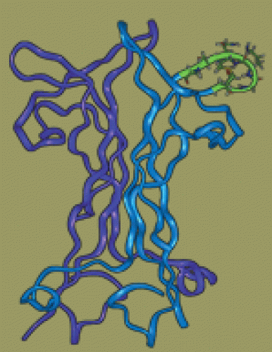
Longo’s team is the first lab to synthesize a small molecule that mimics the effects of a neurotrophin. This “peptidomimetic” is similar in shape to a small but critical part of an active chain, or peptide, of neurotrophin. This similarity in shape allows the synthetic molecule to interact with neurotrophin’s target cells and trigger its normal signaling processes. These small synthetic fragments of neurotrophin are what’s known as “lead compounds” — the first of many steps in drug development.
The next step is to find drug-like chemical compounds that are smaller, more stable, and more potent than the lead compounds. “We’re identifying as many of these small molecule compounds that have the neurotrophin activity as possible,” Longo says. The researchers use a virtual library of synthetic and natural compounds to find those that match the desired size and shape. “In the old days you would have to physically test these compounds one at a time in dishes with neurons,” Longo says. “It would take you decades, because there are millions of compounds in the world.” With the computer library, the researchers can screen the structures of more than 500,000 compounds in only a few hours.
In the lab and in the clinic
Neurons also die as a consequence of AIDS dementia, which is similar to other dementias but has symptoms and mechanisms all its own, including cognitive and motor slowing as well as death of neurons in certain regions of the brain. To understand how HIV damages neurons, Rick Meeker, research professor of neurology, works with an M.D. and a neuropsychologist who treat AIDS patients and evaluate their cognitive function — Colin Hall, professor of neurology, and Kevin Robertson, research associate professor of neurology.
The three researchers study cerebrospinal fluid (CSF) taken from HIV patients to figure out exactly what happens to the virus in the CSF and whether the brain produces its own virus. It’s likely that the body stores HIV in reservoirs because even though many patients have reduced their virus to undetectable levels with antiretroviral drugs, the therapy never completely eliminates the virus. When patients stop medication, the viral infection often returns. So where is the virus hiding? And is the brain one of those hiding places?
t’s pretty clear that neurons themselves aren’t infected by HIV, Meeker says. It could be that HIV infects and activates other cells known as microglia and macrophages, which then produce toxic factors such as cytokines. Using brain cells from an animal model of HIV — cats infected with Feline Immunodeficiency Virus — Meeker has shown in a culture dish that either the virus itself or secretions from the macrophages or microglia will kill neurons. There is still a long list of possibilities as to exactly which secretions could be killing the neurons. But from his work, Meeker suspects that the toxins are disabling or killing the cells by disrupting the balance of calcium inside the cell (see Calcium Overload, page 10).
Hall, Robertson, and Meeker have shown that CSF from human AIDS-dementia patients does contain toxic factors similar to those Meeker has seen in the animal models. Hall and Robertson are collecting data in the clinic to find out whether they can correlate what they see in the patients’ CSF with the patients’ symptoms. Do patients whose CSF contains high levels of virus or toxic factors show increased dementia? In order to show signs of dementia, must patients show a certain strain of virus in their CSF, toxins in their CSF, or both? “We’re trying to figure out if the toxic processes we are seeing in cultured neurons have relevance for the patient,” Meeker says. “This may eventually have some diagnostic or therapeutic utility.”
A new hope
Basic scientists and clinicians alike are optimistic that rapid advances will yield progress in treating neurological disorders. Snider, for instance, is convinced that nerve growth factors will be the key to someday figuring out how to make an injured spinal cord regenerate. And he’s enthusiastic about the future of the field in the hands of young scientists such as new recruits Jay Brenman and Larysa Pevny. “I think that’s where the major breakthrough is going to come from — talented people who are committed to studying a problem and who look at things with a fresh eye,” Snider says.
Longo is also optimistic. Right now, he says, we’re seeing the first signs of hope for treatment of diseases such as Alzheimer’s. “In the past four to eight years, the basic mechanisms related to Alzheimer’s have been elucidated for the first time,” he says. Drugs that may slow the progression of the disease are now in phase one (safety testing) or phase two (efficacy testing) clinical trials. “In some cases, from point of discovery to getting to phase one or phase two trials, the time has been on the order of five or seven years,” Longo says. “That’s dramatic.”
Neurons in Action
Used to be that neurobiology students spent hours at a time performing individual experiments on nerve cells. To see how a nerve cell responded to a drug, for example, they would have to do a new experiment for each change in drug concentration.
Now, thanks to a husband-wife scientist team, students have a more efficient way to run experiments — on CD-ROM. Developed by Ann Stuart, professor of cell and molecular physiology, and her husband John Moore, professor emeritus of neurobiology at Duke University, “Neurons in Action” is an interactive learning tool that simulates laboratory experiments on these cells.
Stuart says the idea for the CD-ROM came from her husband, who along with Michael Hines (now at Yale University) had written a simulator called NEURON, which computational neuroscientists use to model nerve function. Designed for professionals, NEURON is normally too complex for students. “My husband had the idea of coupling the power of a browser with that of NEURON to make tutorials. The browser could then link text to NEURON simulations, customized by us,” Stuart says.
“Neurons in Action” includes 17 tutorials organized into progressive levels of difficulty so that it can be used by undergraduate, graduate, or medical students. Students perform experiments by specifying a variety of conditions such as the neuron’s geometry, channel density, environmental temperature, and ionic environment. A simulator then displays movies of changing voltage patterns at each point throughout a nerve cell.
Since the tutorials are formatted in HTML and displayed by an Internet browser, students can click on the various hyperlinks provided in the text, which will take them to background information as well as original classic papers to help them understand their observations. Included in those papers is a discussion of the origin of the Hodgkin-Huxley equations formulated in the 1950s — and still used today — that describe the electrical signals generated in the nerve cell.
“For scientific subjects that can be described quantitatively, I think this represents the wave of the future,” Stuart says. “It gives students the chance to explore important neurophysiological problems in far greater depth than is offered in conventional textbooks and to correct misconceptions on the spot.”
Cells That Become Us
We start our lives as a cluster of cells, but exactly how we go from a bundle of cells to a bundle of joy remains a mystery. How do our cells know to develop into a liver cell, a brain cell, or a red blood cell? And how do our cells retain this information so that they don’t regenerate into a different type of cell altogether? Larysa Pevny, assistant professor of genetics, is working to understand both aspects of this transformation by examining the function of a subfamily of genes known as SOXB1.
Pevny studies in mice the earliest beginnings of the adult nervous system — a region of cells called the neural plate. The adult mouse nervous system, like our nervous system, is made up of neurons and glial cells. But, Pevny explains, it starts as a sheet of like cells called the epiblast. Epiblast cells receive signals that cause them to form three distinct cell layers — the ectoderm, the mesoderm, and the endoderm. Ectoderm cells eventually form skin, hair, the brain, and the nervous system.
Because these primitive cells are a blank slate waiting for the proper signal to decide their fate within the body, they are used extensively by researchers to study the process of cellular differentiation. For example, it is possible to use retinoic acid (RA), a form of vitamin A, to coax ectodermal cells into differentiating into neural cells. RA is a tool used in the lab, but what are the natural signals for this transformation in the body?
To determine if SOXB1 genes initiate neural plate formation, Pevny and collaborators took advantage of the fact that an engineered cell line known as P19 ectodermal cells differentiates into neural cells when exposed to RA in a culture dish. They asked — instead of adding RA, if you maintain expression of SOX1 (one of the three SOXB1 genes), what will the cells become? The answer is neural cells, indicating that SOX1 can predispose ectodermal cells to become neural cells.
Pevny has also shown that while SOXB1 expression is pervasive very early in development, it is maintained later in development predominately in neural stem cells. This loss of expression is an example of the permanent loss of genetic information that is necessary in order for our bodies to function properly. Blood cells don’t regenerate into liver cells because they eventually stop expressing genes whose protein products are necessary to make such a drastic change.
Pevny uses this permanent segregation of the SOXB1 genes to locate living neural stem cells. For example, bright green fluorescent protein (GFP) can be engineered to be expressed only in cells containing SOX2. Because SOX2 expression is mimicked by the presence of neural stem cells, these genes now mark or define living neural stem cells. GFP allows researchers to visualize the target of interest, in this case SOX2, while the mouse is alive.
Because Pevny’s work helps locate these cells, it is now possible to isolate populations of neural stem cells and examine their differentiation properties in a living system. In the future such techniques may be used in transplant studies. And, with the help of GFP, researchers can now clearly see adult neural stem cells, making it easier to study the effects of alcohol, exercise, or smoking on those cells.
Pevny and others have found that SOXB1 proteins can be used to purify mouse embryonic stem (ES) cells so they are more likely to become neural cells. ES cells have the ability to generate neurons, but when researchers differentiate ES cells in the lab, what results is a variety of cell types, including neurons. Because SOXB1 genes are expressed in neural cells, Pevny has used them to suppress the formation of other cell types. This SOXB1-assisted enrichment has resulted in cell populations that are 80 to 90 percent pure neurons. A source of pure neural cells could one day be used in transplant studies involving diseases such as Alzheimer’s and Parkinson’s.
Competent to Die
What makes a living cell commit “suicide?” Cell death, or apoptosis, sounds ominous, but it’s actually a necessary part of normal cellular activity. Mohanish Deshmukh, assistant professor of cell and developmental biology, works to understand exactly what happens when nerve cells, or neurons, die by apoptosis.
If a cell is damaged, and the damage is too extensive, the cell can activate a self-destruct mechanism. This programmed cell death is vital to the survival of the organism because it prevents the damaged cell from producing other damaged cells. “Apoptosis is also important during early embryonic development,” Deshmukh explains. “The developing embryo generates more neurons than are actually needed to ensure that, if any neurons become lost on the way to their target area or are damaged, the body still has enough to make the proper connections. The body uses apoptosis in this case to eradicate unnecessary neurons.”
In his research, Deshmukh uses “sympathetic neurons,” which are part of the peripheral nervous system. Sympathetic neurons are an excellent model system, he says, largely because of their dependence on a single protein called nerve growth factor (NGF). “If these neurons are placed in culture in the presence of NGF, virtually one hundred percent of these neurons will survive, but if the NGF is removed, one hundred percent will die,” Deshmukh says.
At the molecular level, apoptosis is an intricate web of cellular communication. After a cell activates the programmed cell death pathway, cytochrome c, an important protein for all oxygen-breathing organisms, is released from the mitochondria, the “powerhouse of the cell.” Once released, cytochrome c binds to a group of proteins, which eventually leads to the activation of caspases, which are the actual “executioners” of cell death. Caspases destroy the cell by “chopping up” the DNA and other cellular proteins.
Based on the accepted model, Deshmukh questioned whether the release of cytochrome c was sufficient to activate caspases in neurons. To examine this, he injected cytochrome c into cells and found that caspases became activated. But when he injected cytochrome c into sympathetic neurons, caspases were not activated and the neurons remained alive. “Something in these neurons made them completely resistant to cytochrome c-induced apoptosis,” Deshmukh says. “Normally, when neurons reach the point of cytochrome c release, they have been deprived of NGF for a period of time, so some other event is occurring which makes them more susceptible or more competent to die.” To test this idea, Deshmukh injected cytochrome c into sympathetic neurons after depriving them of NGF, and found that under these conditions, caspases were immediately activated and the neurons died rapidly. This finding indicates that in addition to the release of cytochrome c, neuronal apoptosis requires a novel pathway called the development of competence. “It makes perfect sense that neurons would have evolved a system that has additional checks and balances in place compared to other cell types because neurons have to last the entire life of the organism,” Deshmukh says. “Our lab is interested in understanding the molecular events that make up the development-of-competence pathway and determining whether it is unique to neurons.”
To study this pathway, the Deshmukh lab uses a variety of biochemical and molecular techniques such as intracellular injections, localization of certain proteins using antibodies, and mice that have genes deleted in the pathway that is required for activated caspases. Knowledge gleaned from these experiments may eventually lead to new therapeutic strategies that will prevent neuronal loss after injury or disease. For instance, a group of proteins called inhibitors of apoptotic proteins (IAPs) are the “brakes” that stop caspases. Recent findings from Deshmukh’s lab demonstrate that the development-of-competence pathway somehow removes IAPs and that this removal, along with the release of cytochrome c, leads to neuronal apoptosis. The work is exciting, Deshmukh says, because molecules such as IAPs may have the potential to prevent neurons from dying in stroke, spinal cord injury, and neurodegenerative diseases such as Alzheimer’s, Huntington’s chorea, and Parkinson’s.
Catherine House was formerly a staff contributor for Endeavors.
Tiffany Heady was a student who formerly contributed to Endeavors.
Robin Arnette was a student who formerly contributed to Endeavors.
Neuroscience benefits greatly from advances in genetics and proteomics. The Fall 2002 Endeavors will feature some of the researchers who are making those advances.
A demonstration of “Neurons in Action” can be found at http://neuron.duke.edu/niasite/. The CD-ROM is published by Sinauer Associates.


